Compact Bone Diagram Microscope : Osteon : Compact Bone | Human anatomy drawing, Anatomy ... : Long bone, compact bone and spongy bone.
Compact Bone Diagram Microscope : Osteon : Compact Bone | Human anatomy drawing, Anatomy ... : Long bone, compact bone and spongy bone.. In this type of bone, the lamellae are organised into concentric circles, which surround a vertical haversian canal (which transmits small neurovascular and lymphatic vessels). These are mostly compacted bone with little marrow and include most of the bones in the limbs. The scanning electron microscope (sem) is among the most frequently used instruments for examining bone. 8/30/2019 a) your histology atlas should include a labeled diagram of compact bone hyaline cartilage adipose blood b) each connective tissue will. Bone basics and bone anatomyhave you ever seen fossil remains of dinosaur and ancient human bones in textbooks, television, or in person at a museum?
Each central canal contains blood vessels and nerve fibers surrounded by loose connective tissue. Between the rings of matrix, the bone cells (osteocytes) are located in spaces called lacunae. These bones tend to support weight and. Under the microscope, bone can be divided into two types compact bone forms the outer 'shell' of bone. These are mostly compacted bone with little marrow and include most of the bones in the limbs.
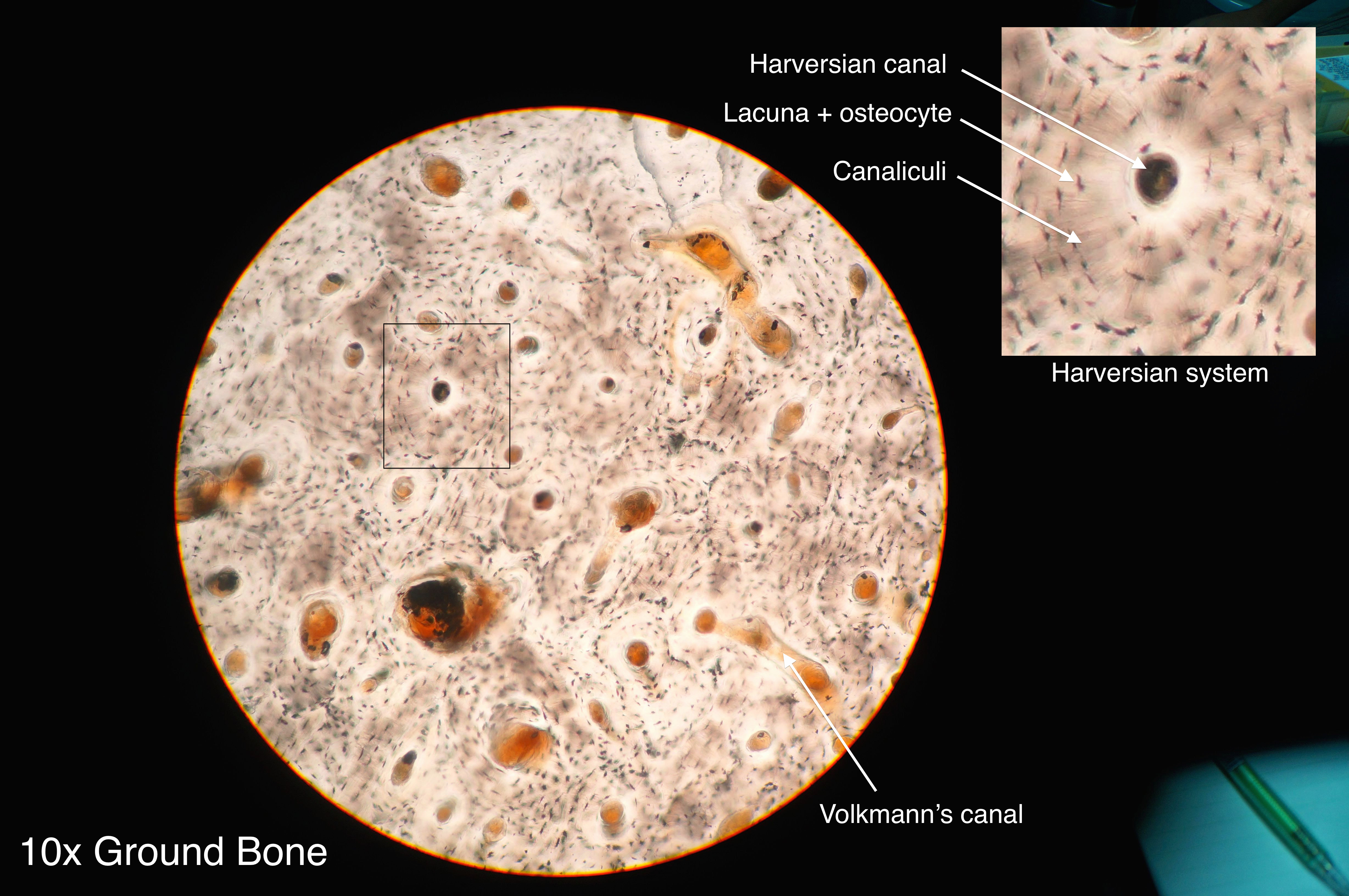
Osteon - Wikiwand from upload.wikimedia.org Bone is an architecturally complex system that constantly undergoes structural and functional optimisation through renewal and repair. Terms in this set (23). The scanning electron microscope (sem) is among the most frequently used instruments for examining bone. Online quiz to learn compact bone microscope slide labeled ; Bone is opaque and must be prepared in the form of thin sections for the brightfield transmission microscopes that are most readily available to amateur microscopists or in educational settings. Draw a labelled diagram of a neuron. A photo taken through a microscope that shows the anatomy of compact bone with a detailed view of an osteon. Each group of concentric circles (each tree) makes up the microscopic structural unit of compact bone called an osteon (this is also called a haversian.
Compact bone, dense bone in which the bony matrix is solidly filled with organic ground substance and inorganic salts, leaving only tiny spaces that contain the osteocytes, or bone cells.
The remainder is spongelike cancellous bone. Virtual microscope slides of cartilage (hyaline, elastic, and fibrocartilage), bone (spongy and compact), and bone development. Label femur diagram handout • review the following terms: What are the 2 main types of bone? Compact and spongy bone with dr. Most think that bone is a dead tissue although compact bone is made up of haversian systems, it is almost solid. Under the microscope, these muscles show alternate light and dark bands or striations when stained appropriately. Compact and cancellous — or compact bone can be found throughout the human skeleton. To bones and help in body movement. Under the microscope dense, compact bone shows a definite and a characteristic pattern of arrangement. Terms in this set (23). The scanning electron microscope (sem) is among the most frequently used instruments for examining bone. Bones protect the various organs of the body, produce red and white blood cells, store minerals.
Compact bone specimens (2x2x36 mm) were harvested from the right metatarsal. Decalcified compact bone at 60x magnification. Each central canal contains blood vessels and nerve fibers surrounded by loose connective tissue. Compact bones make up 80 percent of the human skeleton; The scanning electron microscope (sem) is among the most frequently used instruments for examining bone.
Osteon slide tissue histology | I'm a nurse | Pinterest from s-media-cache-ak0.pinimg.com Compact bone microscope slide labeled learn by taking a quiz; Bone cells are embedded in a hard matrix that is composed of calcium and phosphorus compounds. If you look at compact bone under the microscope, you will observe a highly organized arrangement of concentric circles that look like tree trunks. Electron microscope image of trabecular bone (x100 magnification). Compact bone diagram osteon compact bone ap pinterest anatomy human anatomy and. Begin by identifying the concentric rings of lamellar bone that surround a haversian canal. It's easy to look at these and think of bones as dry, dead sticks in your body, but this couldn't be further from the truth. Compact bones make up 80 percent of the human skeleton;
A diagram of the anatomy of a bone, showing the compact bone.
Each group of concentric circles (each tree) makes up the microscopic structural unit of compact bone called an osteon (this is also called a haversian. The ground substance of bone is arranged in concentrated layers (lamellae) round the small canals which run parallel to the long axis (shaft) of the bone. The basic units of compact bone are called osteons or haversian systems. Illustration about compact bone, also called cortical bone, is the hard, stiff, smooth, thin, white bone tissue that surrounds all bones in the human body. Compact and cancellous — or compact bone can be found throughout the human skeleton. Compact bone specimens (2x2x36 mm) were harvested from the right metatarsal. Bones protect the various organs of the body, produce red and white blood cells, store minerals. Cortical bone forms the extremely hard exterior while cancellous bone fills the interior. Online quiz to learn compact bone microscope slide labeled ; Compact and spongy bone with dr. Samples were cyclically loaded to failure and then histological analyses on each hysteretic diagram, all cycles after the initial monotonic cycle appear pinched and share two points. Bone is an architecturally complex system that constantly undergoes structural and functional optimisation through renewal and repair. A bone is a rigid tissue that constitutes part of the vertebrate skeleton in animals.
Bone is an architecturally complex system that constantly undergoes structural and functional optimisation through renewal and repair. Under the microscope, bone can be divided into two types compact bone forms the outer 'shell' of bone. Draw a labelled diagram of a neuron. Compact bones make up 80 percent of the human skeleton; 8/30/2019 a) your histology atlas should include a labeled diagram of compact bone hyaline cartilage adipose blood b) each connective tissue will.
Microscope Slides Exam 1 - Exercise Science 222a with ... from classconnection.s3.amazonaws.com Quickly memorize the terms, phrases and much more. A bone is a rigid tissue that constitutes part of the vertebrate skeleton in animals. Compact bone diagram osteon compact bone ap pinterest anatomy human anatomy and. The osteon consists of a central canal called the osteonic (haversian) canal, which is surrounded by concentric rings (lamellae) of matrix. Cortical bone forms the extremely hard exterior while cancellous bone fills the interior. What are the 2 main types of bone? If you look at compact bone under the microscope, you will observe a highly organized arrangement of concentric circles that look like tree trunks. Under the microscope, bone can be divided into two types compact bone forms the outer 'shell' of bone.
Compact bones make up 80 percent of the human skeleton;
Draw a labelled diagram of a neuron. After you've studied all the pieces of the compound microscope, it's time to put your brain to the test. The ground substance of bone is arranged in concentrated layers (lamellae) round the small canals which run parallel to the long axis (shaft) of the bone. A cross section of decalcified compact bone is examined under brightfield illumination with the intel qx3 microscope. Virtual microscope slides of cartilage (hyaline, elastic, and fibrocartilage), bone (spongy and compact), and bone development. Next we have a blank microscope diagram. Microscopic structure of a compact bone. Cortical bone is compact bone, while cancellous bone is trabecular and spongy bone. One advantage for the amateur is that the preparation of bone specimens does not require the use of. The osteon consists of a central canal called the osteonic (haversian) canal, which is surrounded by concentric rings (lamellae) of matrix. The basic units of compact bone are called osteons or haversian systems. In this type of bone, the lamellae are organised into concentric circles, which surround a vertical haversian canal (which transmits small neurovascular and lymphatic vessels). Cortical bone forms the extremely hard exterior while cancellous bone fills the interior.
What are the 2 main types of bone? compact bone diagram. Online quiz to learn compact bone microscope slide labeled ;